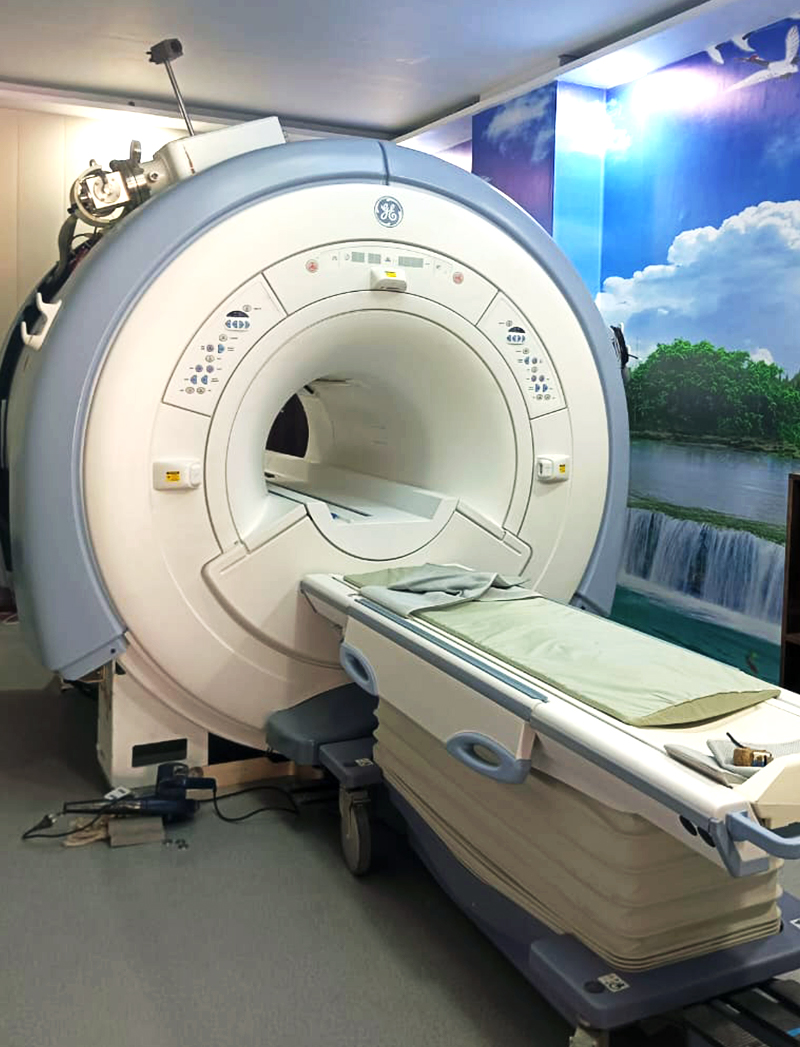
MRI
(Magnetic resonance imaging)
Magnetic resonance imaging (MRI) uses a powerful magnetic field, radio waves to create a picture of the inside of the body. At Royal Scans, we routinely perform MRIs to help evaluate and diagnose numerous conditions.
1.5 Tesla MRI At Royal Scans
At Royal Scans, we routinely perform MRIs to help evaluate and diagnose numerous conditions. Magnetic resonance imaging (MRI) utilizes a powerful magnetic field that generates radio waves to create a picture of the body’s inside. Our department also performs several specialty imaging procedures such as brain spectroscopy, magnetic resonance angiography (MRA), prostate MRI, and venography (MRV). In pregnant women, body MRI may be used to monitor your baby safely.
A wide range of both plain and contrast scans including MRI Head, Foot, Spine, Chest, Ankle Joint, Elbow Joint, Knee, Hip Joint, Leg, Shoulder, Wrist, Abdomen, Pelvis, Orbit, Hand and many others are performed.
What is MRI?
Magnetic resonance imaging (MRI) is a non-invasive test used which is used by radiologists to diagnose medical conditions. MRI uses a powerful magnetic field, radio waves, and a computer to produce images of internal body structures which are in great detail. Detailed images help doctors in finding and detecting diseases.
What are some common applications of the MRI procedure?
MRI imaging of the body is performed to analyse:
- organs of the chest and abdomen—such as the liver, the heart, kidneys, biliary tract, spleen, pancreas, bowel, and adrenal glands.
- Pelvic organs involve the bladder and reproductive organs such as the and prostate glands in males and uterus and ovaries in females.
- Blood vessels
- lymph nodes.
MRI is also used for other medical conditions such as:
- tumours of the pelvis, chest and abdomen.
- abnormalities of the bile ducts and pancreas and diseases of the liver, such as cirrhosis.
- inflammatory bowel disease like, Crohn’s disease
- heart problems including congenital heart disease.
- inflammation of the vessels (vasculitis) and malformations of the blood vessels.
- a foetus in a pregnant woman.
- To find out Orthopaedic conditions & abnormalities
How to Prepare for the Procedure?
Before the Procedure
- You will be told to wear a hospital gown.
- Unless you are told otherwise, you should take food and medications as usual.
- Tell your radiologists if you have undergone recent surgery. Gadolinium contrast is used for MRI, which is considered safe for patients with kidney disease. Pregnant women also should tell radiologists if they are pregnant.
- If you have claustrophobia or anxiety, the doctor may prescribe a mild sedative prior to your exam.
- Prior to the scan, leave all the jewellery and other accessories at home.
- Tell the radiologists if you have medical or electronic devices in your body such as cochlear (ear) implants, types of clips used for brain aneurysms, metal coils placed within blood vessels, older cardiac defibrillators and pacemakers.
During the Procedure
- MRI does not use radiation, and hence, it causes no chemical changes in the tissues.
- You will be placed on the moveable exam table, and straps and bolsters are used to help you maintain the position. While the images are being taken, it is important that you remain perfectly still.
- Hydrogen atoms naturally exist within the body re-align with the radio waves, and the scanner captures this energy to develop a picture using this information.
- The computer processes the signals and helps in creating a series of images. The radiologists study the images from different angles.
- After the exam, you will be asked to wait while the radiologists check the images.
- The entire exam is usually completed in 30 to 50 minutes, depending on the type of exam and equipment used.
After the Procedure
- Technologists or a doctor will interpret medical exams. Your reports will be shared with you.
- Sometimes, the patient has to take follow-up exams because of an abnormality that may need further evaluation with additional views or a special imaging technique.
- Also, a follow-up exam tells whether the treatment being used to treat the medical condition is working or not.
Are there any risks involved?
- When appropriate safety guidelines are followed, the MRI exam poses almost no risk to the average patient.
- There are no risks from the strong magnetic field. However, it may result in distorted images or malfunction of the implanted medical devices.

1.5 Tesla MRI At Royal Scans
At Royal Scans, we are proud to offer state-of-the-art 1.5 Tesla MRI imaging services. Our advanced MRI technology provides exceptional image quality, enabling accurate and detailed diagnostic information for our patients.
When you choose Royal Scans for your 1.5 Tesla MRI, you can trust that you are receiving top-notch imaging services in a welcoming and patient-centered environment. We are committed to providing you with the highest quality of care, ensuring that your diagnostic journey is seamless and efficient.
Plan For Your Visit
Reliable Results
“Count on our labs for reliable results you can trust, providing you with the information you need for informed healthcare decisions.”
Fully Trained Staff
“Our labs boast a team of highly trained staff, ensuring the utmost precision and professionalism in every test we perform.”
Patient Care
“Our dedicated customer care team is here to provide you with exceptional service and support throughout your healthcare journey.”
